
Figure 2.
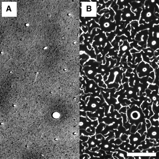
Quality of individual staining for CO activity and blood vessels in double-stained specimen, here a 60-μm-thick tangential section from a squirrel monkey. A, A 50× magnified bright-field micrograph from a similar section as in Figure 1 showing some CO blobs. B, Fluorescence micrograph from the same area of the same section as in Figure 3A, showing the blood vessels stained with a primary antibody against collagen type IV and a secondary antibody labeled with Cy3. Scale bar, 200 μm.