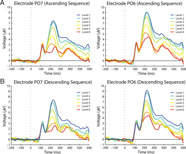
Figure 4.
Waveform P2 component (main experiment). Left (PO7) and right (PO6) parieto-occipital electrodes in which P2 modulations can be clearly distinguished. A shows amplitude modulations per degradation level during the ascending sequence. B shows amplitude modulations during the descending sequence.

