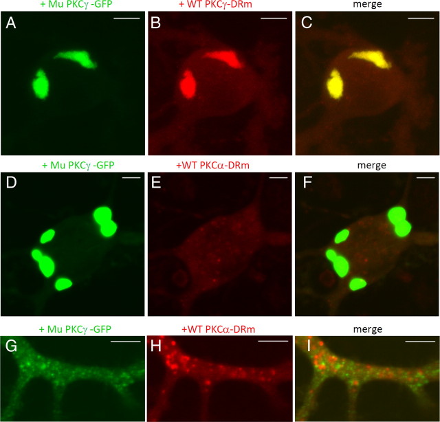
Figure 6.
Colocalization and aggregation of wild-type PKCγ-DRm, but not wild-type PKCα-DRm, with mutant PKCγ-GFP. A–C, Coexpression of mutant (Mu) PKCγ-GFP with wild-type (WT) PKCγ-DRm in cultured PCs. D–I, Coexpression of mutant (Mu) PKCγ-GFP with wild-type (WT) PKCα-DRm in cultured PCs. Fluorescence images of GFP (A, D, G), DsRed monomer (B, E, H), and the superimposed images (C, F, I) in the soma (A–F) and the dendritic shaft (G–I). Although PKCα-DRm appears to form small aggregates in the dendrite (H), they are in lysosomes; thus, they are not aggregates. Similar dots can be observed when only the DsRed monomer was expressed in cultured PCs. Scale bars: A–F, 10 μm; G–I, 5 μm.

