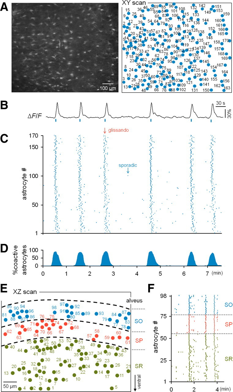
Figure 2.

Calcium activities of astrocytes that were synchronized throughout the dorsal hippocampus. A, Two-photon image of an XY section of the stratum oriens in a fluo-4-loaded hippocampus (left) and the identified location of the somata of individual astrocytes. B, Typical trace of the calcium fluorescence intensity of a single astrocyte. C, Rastergram of the astrocytic activities recorded in A. Each dot indicates the timing of a single calcium event. Synchronous activities (glissandi) were evident in the background of sparse activities (sporadic). D, Percentage of active astrocytes at a given time (10 s bin). E, Map of astrocytes in an XZ section across the stratum oriens (SO), stratum pyramidale (SP), and stratum radiatum (SR) in the hippocampus. F, Rastergram of the astrocytes shown in E.
