
Figure 3.
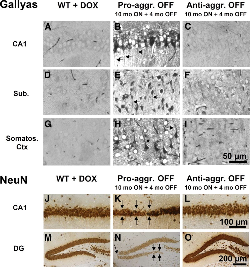
Histology of pro-aggregant and anti-aggregant TauRD mice at 10 months switched ON plus 4 months OFF compared with wild-type mice. Top, Gallyas silver staining for Tau aggregates. Bottom, NeuN staining for neurons. Images B, E, and H illustrate Gallyas-positive neurons in the CA1 region, subiculum, and somatosensoric cortex of pro-aggregant OFF mice compared with wild-type mice (A, D, G) and anti-aggregant OFF mice (C, F, I). Note the missorting of Tau into the apical dendrites of the neurons in pro-aggregant OFF mice (B, E, H, arrows). Images K and N show the neuronal loss in the CA1 region and DG of pro-aggregant OFF mice (arrows) compared with wild-type (J, M) and anti-aggregant OFF (L, O) mice. Note shrinkage of entire pyramidal cell layer in pro-aggregant OFF mice (K) in contrast to wild-type (J) and anti-aggregant OFF (L) mice. Pro-aggr., Pro-aggregant; Anti-aggr., anti-aggregant; WT, wild type; DOX, doxycycline; Sub., subiculum; Somatos. Ctx, somatosensoric cortex. Scale bars: A–I, 50 μm; J–L, 100 μm; M–O, 200 μm.