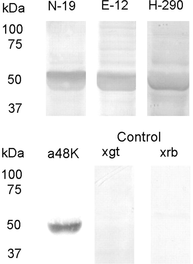
Figure 1.
Western blot of scallop retinal tissue. Retinal proteins separated on SDS-PAGE (12%) were electrotransferred to nitrocellulose membranes and incubated (1:1000) with several antibodies raised against vertebrate arrestins, followed by secondary Abs conjugated to alkaline phosphatase, and development. All antibodies produced a band of similar apparent molecular mass, ≈50 kDa. Omission of the primary antibodies and incubation with either anti-goat (×gt) or anti-rabbit (×rb) alkaline phosphatase-conjugated antibodies did not produce any signal.

