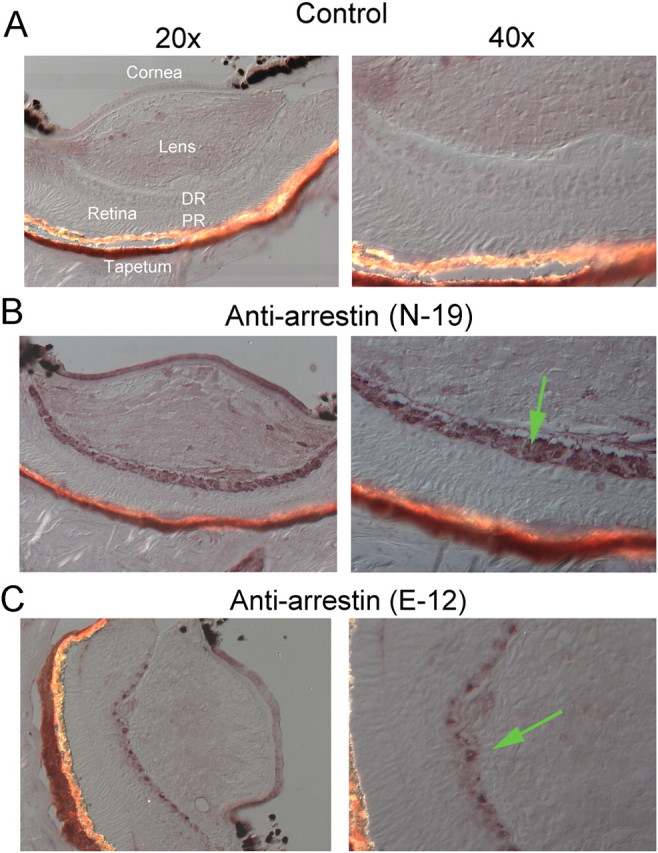
Figure 2.

Immunolocalization of arrestin in Pecten eye. A, Nomarski micrographs of a control 12-μm-thick cryosection through a whole scallop eye, fixed in paraformaldehyde. The section was incubated only with secondary antibodies, and is displayed at two magnifications (RD, distal retina; PR, proximal retina). B, Section treated with one of the anti-arrestin antibodies (N-19); a distinct immunostaining pattern is observed, which is confined to the distal retinal layer, where the ciliary, hyperpolarizing photoreceptors are located (arrow in lower right panel). C, Staining by the E-12 antibody was also segregated to the distal retina.
