
Figure 2.
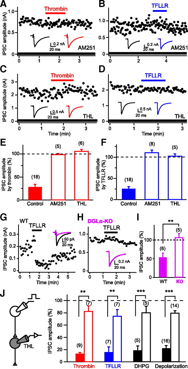
Involvement of endocannabinoid signaling in PAR1-induced IPSC suppression. A–D, Representative data showing effects of AM251 (0.3 μm; A, B) and THL (5 μm; C, D) on IPSC suppression induced by thrombin (1 U/ml) or TFLLR (10 μm). Representative IPSC traces acquired before (left) and during (right) application of thrombin or TFLLR are shown in insets. E, F, Summary graph showing normalized IPSC amplitudes during application of thrombin (E) or TFLLR (F) in the presence of AM251 and THL. G–I, TFLLR-induced IPSC suppression is absent in DGLα-knock-out (KO) mice. Data are illustrated similarly to Figure 1J–L. J, Intracellular application of THL (10 μm) to a postsynaptic neuron greatly reduced IPSC suppressions by thrombin (1 U/ml), TFLLR (10 μm), DHPG (20 μm), and postsynaptic depolarization (5 s) to the similar extents. Filled and open columns represent normalized IPSC amplitudes for control and THL-injected neurons. Numbers in parentheses represent the number of tested cells. WT, Wild-type. **p < 0.01, ***p < 0.001. All data are presented as mean ± SEM.