Figure 6.
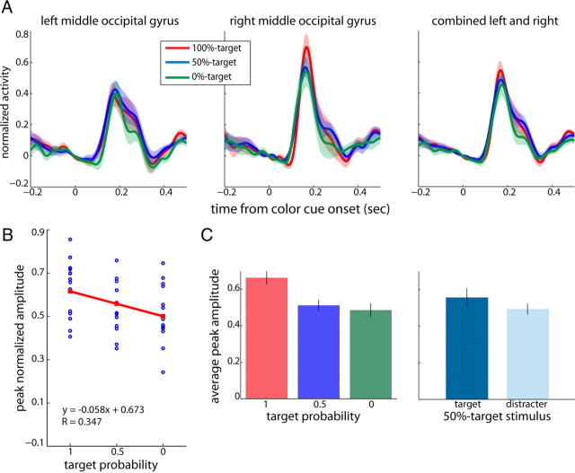
Target selection: extrastriate visual cortex. A, Average virtual sensors for left and right middle occipital gyri and combined across both hemispheres (from left to right) as a function of time from color cue onset. The different colors represent responses to contralateral stimuli of varying target probabilities (red: 0, blue: 0.5, green: 0). Shaded areas represent SEM. B, Corresponding peak normalized amplitudes for all subjects (blue dots) as a function of target probability for the combined dataset. Regression lines are indicated in red. C, Average peak amplitudes for the combined dataset (left and right middle occipital gyri). Left, Average peak amplitudes as a function of target probability. Right, Average peak amplitudes for the 50%-target stimulus when presented as a target (dark blue) or distracter (light blue). Black lines are SEM.

