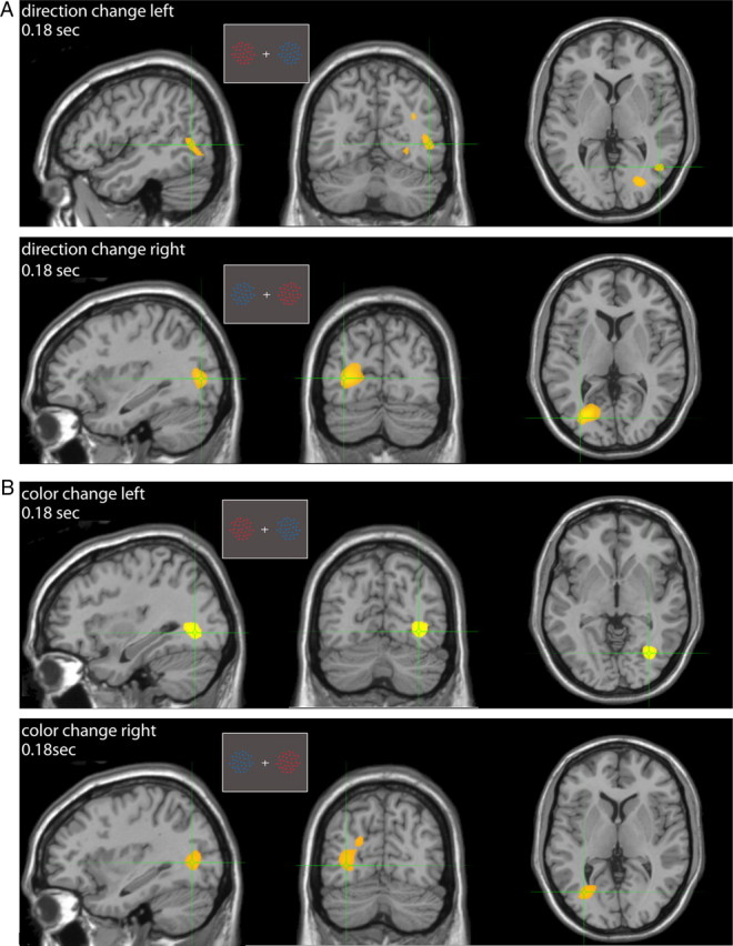
Figure 8.

Sustained attention: group erSAM analysis. A, Sagittal, coronal, and axial views of brain regions maximally activated (yellow) 180 ms after the transient direction change in the target when positioned on the left (top) or right (bottom). B, The same as in A for transient color changes. Insets represent example stimulus configurations.
