Figure 1.
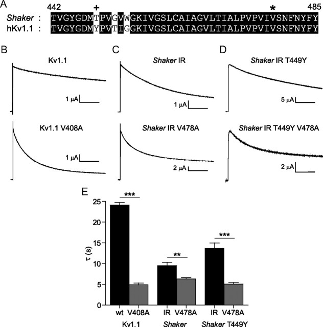
A valine to alanine mutation accelerates current decay in Kv1.1 and Shaker channels. A, An alignment of the selectivity filter and S6 pore lining regions in Shaker and Kv1.1 demonstrates 93% amino acid conservation across the region shown. * denotes site of EA-1 mutant V408A (in Kv1.1) and the equivalent mutant V478A in Shaker. + denotes a Shaker site “T449” (Kv1.1 Y379), a poorly conserved site involved in modulation of C-type inactivation. B–D, Currents during pulses to +60 mV from a holding potential of −80 mV are shown for Kv1.1 and Kv1.1 V408A (B), Shaker IR and Shaker IR V478A (C), and Shaker IR T449Y and Shaker IR T449Y V478A (D). E, Mean time constants from fits to traces in B–D. Measured mean τ values were as follows: 24.1 ± 0.6 s (Kv1.1 wt), 4.89 ± 0.42 s (Kv1.1 V408A); 9.52 ± 0.76 s (Shaker IR), 6.30 ± 0.30 s (Shaker IR V478A); 13.7 ± 1.3 s (Shaker IR T449Y), 5.08 ± 0.35 s (Shaker IR T449Y V478A). Comparisons were made using one-way ANOVA with Bonferroni post hoc tests for significance, **p < 0.01 or ***p < 0.001. Horizontal scale bars denote 5 s.

