
Figure 4.
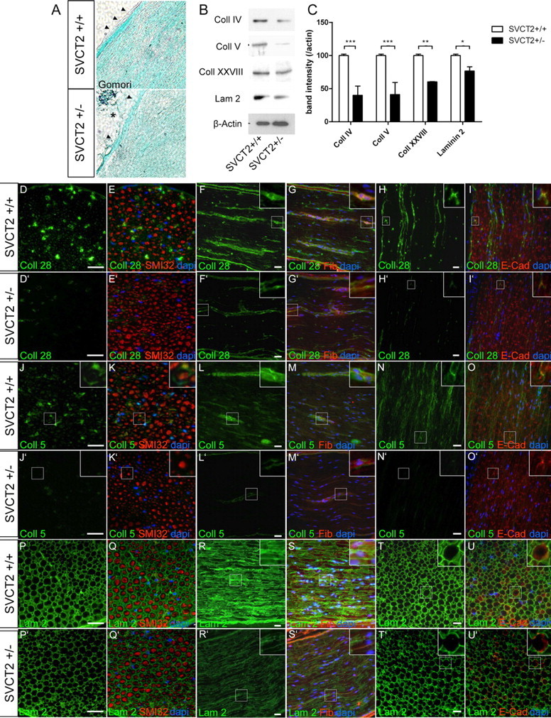
Formation of collagen (Coll)-containing and laminin (Lam)-containing extracellular matrix is reduced in SVCT2+/− peripheral nerves. To investigate the extracellular matrix of SVCT2+/− peripheral nerve tissue, we studied sciatic nerves by light microscopy, immunofluorescence microscopy, and Western blots. A, Light microscopic imaging following Gomori's staining showed only mild changes in SVCT2+/− nerves (bottom) compared to wild-type nerves (top). SVCT2+/− nerves showed slightly disorganized collagen fibrils (green), particularly in the epineurium (arrowheads), which appeared partially disrupted (bottom image, asterisk). B, Western blots of sciatic nerve lysates showed reduced band intensities for collagen IV, V, and XXVIII and for laminin-2. Actin was used as a marker for protein loading. C, Densitometric analysis of Western blot bands confirmed significant downregulation of collagens IV, V, and XXVII, as well as laminin-2 (***p < 0.001, **p < 0.01, *p < 0.05, n = 3). D–U′, Immunohistochemical analysis of collagen types XXVIII, V, and laminin-2 (green) and merged images with counterstaining using neurofilament antibodies (SMI32, red), fibronectin (Fib) antibodies (red), and E-cadherin (E-Cad) antibodies (red), as well as DAPI (blue) on cryosections of sciatic nerves from SVCT2+/− and wild-type mice, are shown. D–I′, Immunostainings of the peripheral nerve-specific collagen type XXVIII showed reduced expression in SVCT2+/− nerves (D–I) compared to wild-type nerves (D′–I′). In the wild-type nerves, collagen XXVIII was localized in the extracellular matrix as shown by double-labeling with fibronectin (F, G, insets) and in the paranodal region of nodes of Ranvier, as shown by double-labeling with E-cadherin (H, I, insets). In both locations, collagen XXVIII was reduced in SVCT2+/− mice (F′, G′, D′, I′, insets). J–O′, Immunohistochemistry of collagen type V showed strong expression in wild-type nerves (J–O), which was markedly reduced in SVCT2+/− animals (J′–O′). Collagen V in the extracellular matrix was strongly reduced (compare M to M′, insets). Accumulation of collagen V at nodes of Ranvier was also reduced in SVCT2+/− nerves (compare O to O′, insets). P–U′, Stainings with antibodies to laminin-2 showed a relative reduction in intensity in SVCT2+/− nerves (P′–U′) compared to wild-types (P–U). Scale bars: 20 μm.