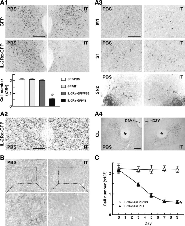
Figure 3.

Selective elimination of PF neurons innervating the dorsolateral striatum. A, Impact of IT treatment on cells transduced retrogradely by the HiRet vector. Mice (n = 4 for each group) received a bilateral injection of the HiRet–IL-2Rα–GFP or HiRet–GFP vector into the dorsolateral striatum, and 5 weeks later they were treated with IT or PBS by injection into the unilateral PF. On day 7 after the treatment, brains were processed for histological examinations. GFP immunohistochemistry of sections through the PF (A1). The top shows representative images of GFP immunohistochemistry, and the bottom shows the number of GFP-positive cells per section. *p < 0.001 compared with each of the HiRet–GFP/PBS, HiRet–GFP/IT, and HiRet–IL-2Rα–GFP/PBS groups (Bonferroni's test). Cresyl violet staining of PF sections prepared from the HiRet–IL-2Rα–GFP-injected mice (A2). GFP immunohistochemistry of M1, S1, and SNc sections prepared from the HiRet–IL-2Rα–GFP-injected mice (A3). GFP immunostaining of CL sections prepared from the same animals (A4). B, Anterograde tracing of axons arising from the PF. The HiRet–IL-2Rα–GFP vector-injected animals treated with IT or PBS by unilateral injection into the PF were used for BDA injection into the PF, and 7 d later their brains were processed. Sections through the dorsolateral striatum were prepared and stained for BDA. Bottom images are magnified views of the dotted rectangles in the top images. C, Time course of cell elimination. Mice (n = 4 for each group) were given a bilateral injection of the HiRet–IL-2Rα–GFP vector and treated unilaterally with IT or PBS into the PF. The brains were processed on different days after the treatment and stained by GFP immunohistochemistry. The number of GFP-positive cells per section was plotted. D3V, Dorsal third ventricle; fr, fasciculus retroflexus. *p < 0.001 compared with the HiRet–IL-2Rα–GFP/PBS group, Bonferroni's test. Scale bar, 200 μm.
