
Figure 11.
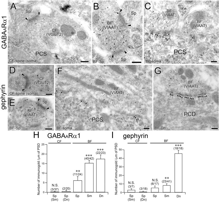
Postembedding immunogold for GABAARα1 and gephyrin in PC synapses at P12. A–C, Double-labeling immunogold for GABAARα1 (ϕ = 10 nm) and VGluT2 (ϕ = 15 nm) or VIAAT (ϕ = 15 nm) at CF–spine (A), BF–spine (B), and BF–soma (C) synapses. Note the GABAARα1 labeling at the asymmetrical contact of the BF–spine synapse (C). D–G, Double-labeling immunogold for gephyrin (ϕ = 10 nm) and VIAAT (ϕ = 15 nm) or VGluT2 (ϕ = 15 nm) at CF–spine (D), BF–spine (E), BF–soma (F), and BF–dendrite (G) synapses. Note the GABAARα1 labeling at the asymmetrical contact of the BF–spine synapse (C). Edges of the PSD and immunogold particles for GABAARα1 and gephyrin are indicated by arrowheads and arrows, respectively. H, I, Summary bar graphs comparing the labeling density for GABAARα1 (H) at CF–spine, BF–spine, BF–soma, and BF–dendrite synapses. The number of immunogold particles (mean ± SEM) for GABAARα1 per synapse is 0.1 ± 0.0, 0.2 ± 0.1, 1.7 ± 0.5, 3.7 ± 0.4, and 6.0 ± 0.8 in CF–somatic spine, CF–dendritic spine, BF–spine, BF–soma, and BF–dendrite synapses, respectively. The number of immunogold particles for gephyrin per synapse is 0.6 ± 0.3, 0.3 ± 0.2, 1.4 ± 0.6, 2.4 ± 0.6, and 21.4 ± 3.6 in CF–somatic spine, CF–dendritic spine, BF–spine, BF–soma, and BF–dendrite synapses, respectively. N.S., p > 0.05; **p < 0.01; ***p < 0.001 (Mann–Whitney U test). Scale bars: 200 nm. PCS, Purkinje cell soma.