Figure 8.
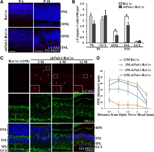
Retinas lacking PALS1 undergo persistent programmed cell death at maturation of the retina. A, Immunohistochemistry for c-CAS3 on representative sections of shPals1-RxCre and RxCre retinas at P6 and P18. Apoptotic cells were stained with c-CAS3 antibodies (red). Nuclear DNA was counterstained with TO-PRO-3 (blue). Scale bar, 50 μm. B, Histogram depicting the number of c-CAS3-positive cells in shPals1-RxCre and control retinas at P6 and P18. Asterisk (*) indicates significant difference compared to the control (Student's t test, p < 0.0017 for P18 OLM, p < 0.0029 for P18 INL). Error bars indicate +SEM. C, Immunohistochemistry for apoptotic cells and bipolar cells on representative sections of shPals1-RxCre and control RxCre retinas at 12 months of age. Apoptotic cells were stained with c-CAS3 antibodies (red, arrowheads), and bipolar cells with anti-PKCα (green). Nuclear DNA was counterstained with TO-PRO-3 (blue). Dashed quadrants in the upper panels indicate examples of a c-CAS3-positive cell, and are shown enlarged in the panel down on the left (scale bar, 10 μm). The number of photoreceptor and bipolar cells decreased over time. Scale bar, 50 μm. D, Quantification of the outer nuclear layer thickness. ONL thickness measured every 500 μm from the optic nerve to an area near the peripheral edge of retinal sections in control (12M) and shPals1-RxCre (3, 6, 12M). Error bars indicate ±SEM.

