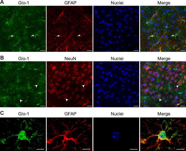
Figure 3.
High expression of Glo-1 in astrocytes of the mouse cerebral cortex. Coronal sections of mouse brain were immunostained with Glo-1 and with the astrocytic marker GFAP (A, C) or the neuronal marker NeuN (B). Glo-1 immunostaining was strongest in astrocytes (A, arrows), but was also present in neurons at lower levels (B, arrowheads). C, Scaled-up image of a single astrocyte in the mouse cerebral cortex shows that Glo-1 immunoreactivity is located in the cell body and processes along the GFAP+ filaments. Representative images from one of three animals are shown. Nuclei are stained using Hoechst. Scale bars: 15 μm (A, B) and 7 μm (C).

