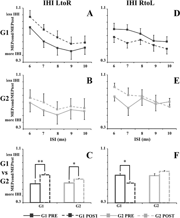
Figure 2.
IHI from LtoR and RtoL hemispheres in G1 (dark gray square) and G2 (light gray square) before (PRE, solid line) and after (POST, dashed line) immobilization. A (G1) and B (G2) illustrate LtoR IHI expressed as the ratio between the mean peak-to-peak MEP amplitude in conditioned versus unconditioned trials (MEPcond/MEPtest on the ordinate), whereas D (G1) and E (G2) illustrate the RtoL IHI. On the abscissa, the ISIs (6, 7, 8, 9, and 10 ms) are shown. On C and F, the mean values of IHI in G1 (dark gray) and G2 (light gray) before (solid line) and after (dashed line) immobilization are shown. Data are represented as mean values ± SE. In A, B, D, and E, asterisks indicate significant difference between pre and post values when interaction of time × ISI was statistically significant. In C and F, asterisks indicate the main effect of time. *p < 0.05, **p < 0.01.

