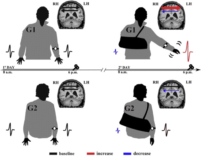
Figure 3.
A schematic view of the experimental design and main results. All participants in G1 and G2 were instructed not to use the dominant (right) hand for 10 h, wearing a soft bandage. The use of the left, not-immobilized arm was monitored by means of an actigraph for 10 h during the period of immobilization (2° day) and 1 d before (1° day). MEP size decreased in the left hemisphere (reduced MEP in blue, compare with black one) in both G1 and G2, whereas it increased in the right hemisphere only in G1 (increased MEP in red, compare with black one). LtoR IHI was reduced in both G1 and G2 (thin arrow in blue, compare with black one), whereas RtoL IHI was deeper only in G1(thick arrow in red, compare with black one).

