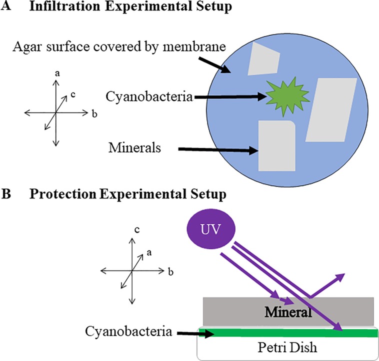
Fig 1. Experimental setup schematics.
A). Infiltration experimental setup. Leptolyngbya cells were inoculated on the surface of a dialysis membrane covered agar, and three mineral flakes, each at approximately 1 cm (length) x 1 cm (width) x 0.25 (thickness) cm in size, were placed nearby. Cells were allowed to grow into the mica sheets for 2 weeks. B). Protection experimental setup. Leptolyngbya cells were inoculated on the surface of membrane-covered agar surface in a petri dish without mineral flakes. After growth for two weeks, the grown biofilm in petri dish was placed under a mica mineral flake and was exposed to UVR.

