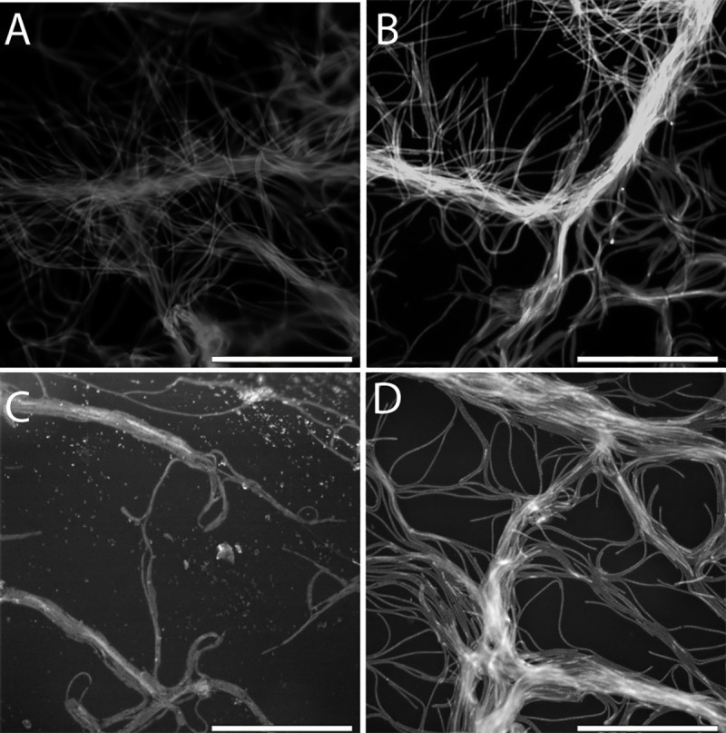
Fig 6. Fluorescent images showing UVB-induced chlorophyll a degradation as a function of mineral type and thickness.

Under thin sheets of phlogopite and biotite (A and C, respectively, both at 0.1 mm), chlorophyll a was partially destroyed, as indicated by the low fluorescent intensity of the individual filaments. Under thicker phlogopite and biotite sheets (B at 3.1 mm and D at 3.8 mm, respectively), chlorophyll a was largely preserved as indicated by the higher signal intensity. Individual cells are clearly visible. There is little difference in chlorophyll a degradation between phlogopite (A and B) and biotite (C and D). For muscovite, even at a small thickness, chlorophyll a was completely photo-bleached (images not shown).
