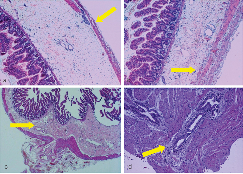
Fig. 2.

(a, b) Microphotograph of CSID (Patient 4) with thin external muscle layer (hematoxylin and eosin ×40) and dilated lymphatic vessels of the submucosa (hematoxylin and eosin staining, ×40 original magnification); (c) microphotograph of CSID (Patient 3) showing sclerosis of the submucosa (hematoxylin and eosin staining, ×20 original magnification); and (d) microphotograph of the “pyloric tumor” showing hypertrophic muscle layers with some scattered glandular proliferations corresponding to a gastric adenomyoma of the pyloric region (hematoxylin and eosin staining, ×100 original magnification). The yellow arrows in (a) and (b) point to the very thin muscularis propria, while the yellow arrows in (c) and (d) point to the sclerosis of the submucosa and to the gastric adenomyoma of the pylorus, respectively. The gastric adenomyoma of the pylorus is characterized by glandular structures lined by cuboidal to columnar epithelium surrounded by hypertrophic smooth muscle bundles by histological examination. CSID, congenital segmental intestinal dilatation.
