Figure 3.
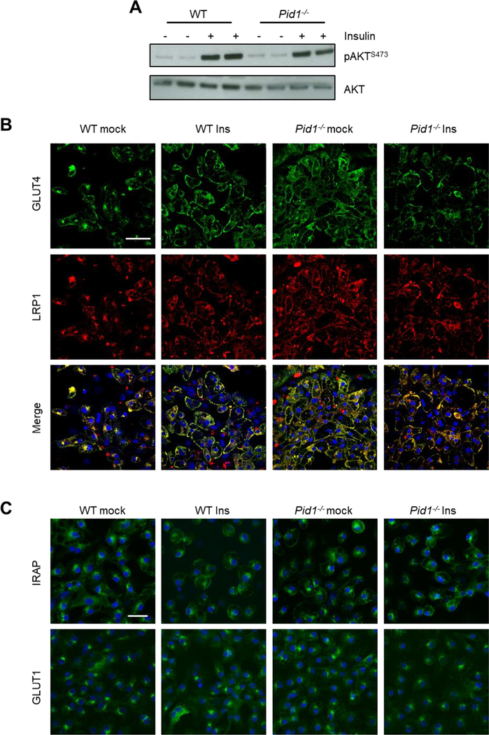
Effect of PID1-deficiency on insulin signaling and localization of LRP1 and GLUT4 in primary brown adipocytes. (A) Insulin signaling was determined by Western blot analysis of AKT phosphorylation at position Ser473. For this purpose, differentiated brown adipocytes prepared from wild type (WT) and Pid1−/− mice were analysed under basal and insulin-stimulated conditions. (B) Immunofluorescence analysis of GLUT4 (green) and LRP1 (red) in WT and Pid1−/− primary brown adipocytes under basal and insulin-stimulated conditions. Yellow fluorescence indicates co-localization. (C) Immunofluorescence analysis of IRAP and GLUT1 in WT and Pid1−/− primary brown adipocytes under basal and insulin-stimulated conditions. Nuclei are stained with DAPI (blue; scale bar in (B) = 20 μm; scale bar in (C) = 25 μm).

