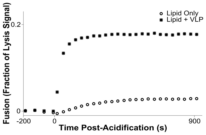
Figure 3. Kinetic assay for hemifusion between influenza VLPs and 5% DiD liposomes.
Vesicles and VLPs are incubated in a 37 C plate reader, and fusion is stimulated by acidification to pH 5.1 at time = 0 s. The acidification results in hemifusion between VLPs and liposomes, dequenching the dye as it is distributed into a larger lipid pool.

