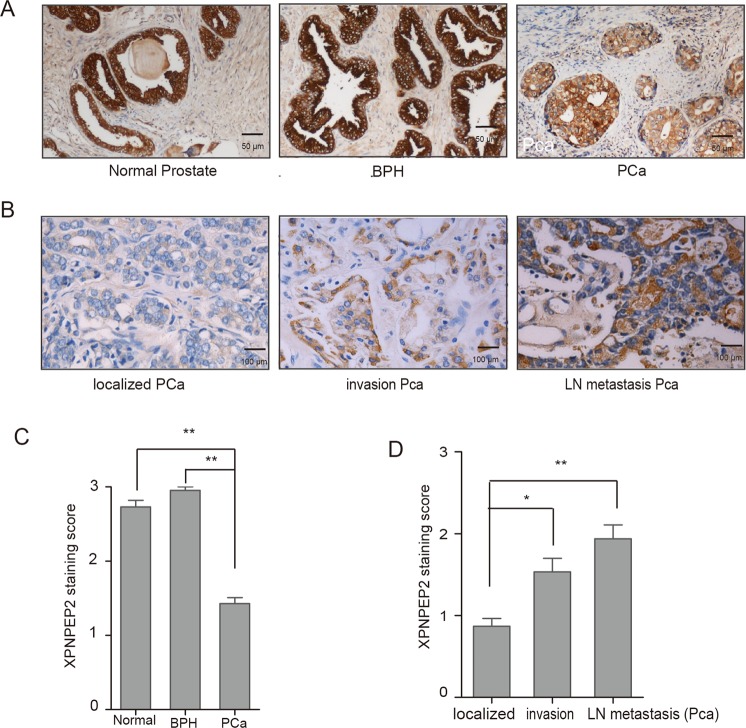
Figure 1.
Immunostaining of XPNPEP2 expression in human prostate tissue. (A) and (C) A prostate cancer tissue microarray PR1921 and 30 BPH tissues were employed for staining with anti-XPNPEP2, the representative figures were shown (A), and XPNPEP2 immunostaining scores was presented (C). Scale bar, 50 um. **p < 0.01. (B) and (D) XPNPEP2 expression in Pca subdivided into localized, locally invasive and LN-metastatic Pca was also analyzed (B, D). Scale bar, 100 um. *p < 0.05, **p < 0.01.

