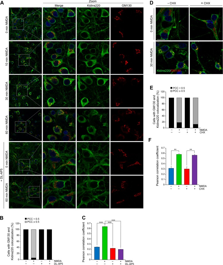
Fig. 3. Excitotoxicity induces Kidins220 translocation to the Golgi apparatus.
a Coimmunostaining of Kidins220 and the Golgi apparatus marker GM130 in neurons treated with NMDA for the indicated times in the absence or presence of the NMDAR antagonist DL-AP5 (200 μM, 60 min). Nuclei were stained with DAPI. Merge confocal microscopy images and magnifications corresponding to the boxed areas are depicted. Scale bar: 10 μm. b, c Percentage of cells displaying colocalization of Kidins220 and GM130 and mean Pearson correlation coefficient (PCC) values, respectively, after 60 min of NMDA treatment, calculated in single optical sections obtained from the different conditions. d Merged magnified images of Kidins220 and GM130 coimmunofluorescence in neurons preincubated with cycloheximide (CHX; 200 μM, 240 min) before NMDA stimulation for 60 min. e, f Percentage of cells displaying colocalization of Kidins220 and GM130 and mean PCC values were calculated as above. Data shown are the means ± s.e.m of three independent experiments after counting n = 20–40 neurons. **p < 0.01, ***p < 0.001, Student’s t-test

