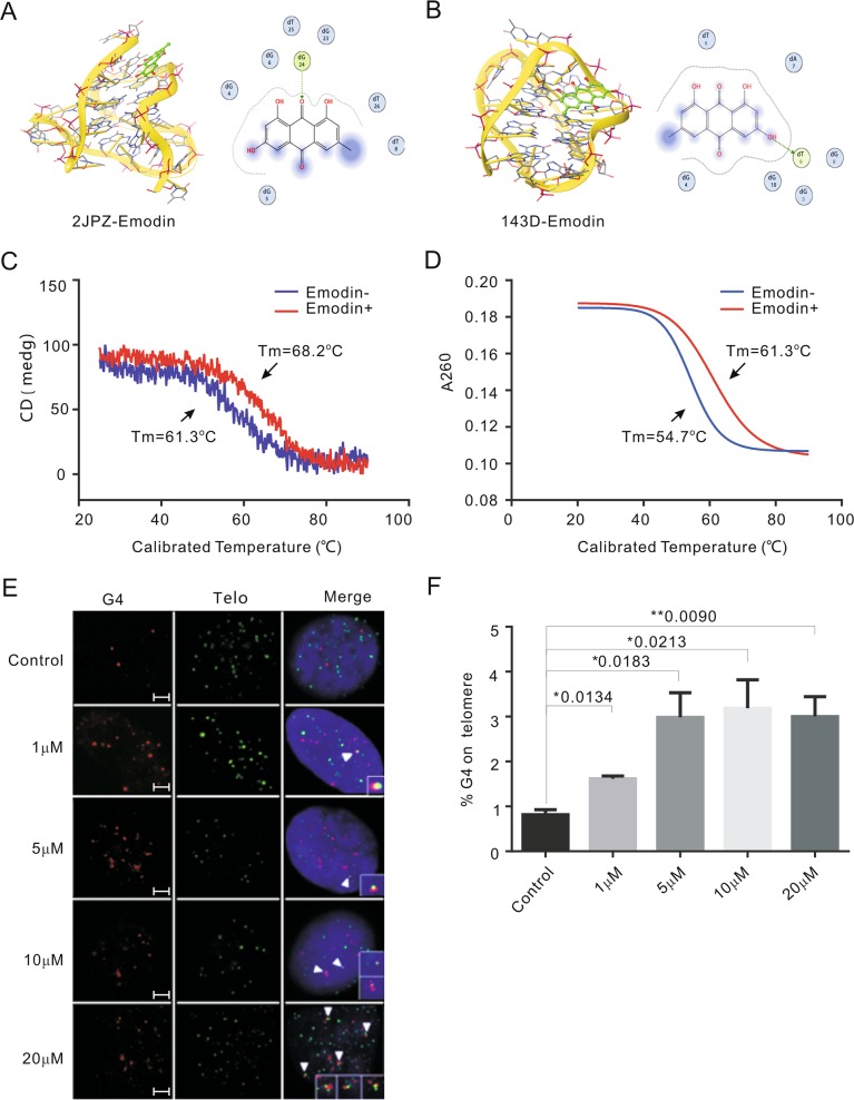
Fig. 1. Emodin stablizes the formation of G4 in vitro.
a, b A molecular docking complex of emodin with hybridized and antiparallel G4. c Circular dichroism melting spectrum reflected the G4 stability with or without emodin treatment by the melting temperature (Tm). d The UV melting reflected the G4 stability with or without emodin treatment by the melting temperature (Tm). e Representative images showing the co-localization of G4 and telomere for the control (DMSO) and emodin-treated HeLa cells (1, 5, 10, and 20 μM for 48 h). The G4 was detected by G4 antibody BG4 (red) and telomere was detected by telomeric FITC-conjugated PNA probes (green). Nuclei were stained with DAPI (blue). White arrowheads in merged images indicate co-localized of G4 and telomere. Scale bars are 2 μm. f Graph shows the percentage of G4 on telomere upon emodin treatment. Data are shown as mean ± SD. More than 100 nuclei were analyzed in three independent experiments

