
Figure 6: p53-null embryos are less sensitive to 5-aza-induced LRA.
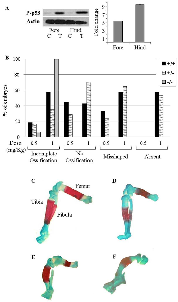
(A) Representative Western blot of phosphorylated p53 in 5-aza treated (‘T’) mice hind limbs destined to be malformed and resistant forelimbs and their controls (‘C’). Quantification was performed in the hind and forelimbs relative to their controls and the Actin levels. (B) Percentage of the different p53 genotypes’ fetuses suffering from the indicated anomalies following 5-aza treatment on GD 10. (B-E) Alizarin red and Alcian Blue staining of cartilage and bone. Limbs were examined on GD 18. (C) Normal long bones: Femur, Tibia and Fibula. (D-F) Representative long bones anomalies: (D) Incomplete ossification of all long bones; (E) All long bones are misshaped, incomplete ossification of Femur and no ossification of the Fibula; (F) Incomplete ossification of the Tibia while the Femur and Fibula are absent.