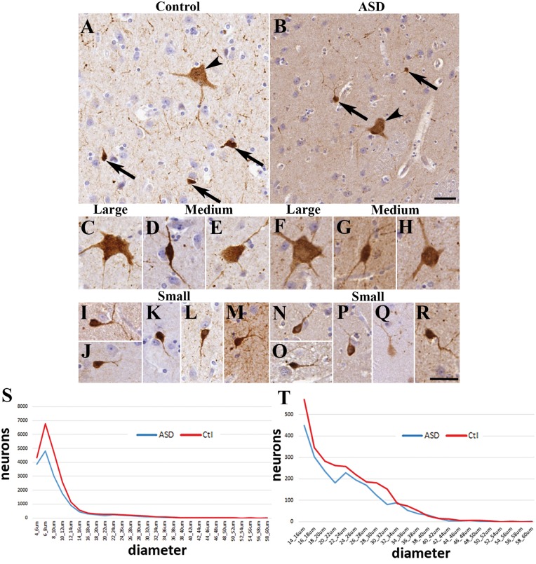
Figure 1.
The morphology of calretinin+ neurons in the caudate nucleus was similar in controls and autism spectrum disorder. Images were taken from four controls and four patients with autism. (A) Representative field from controls and (B) from autism spectrum disorder (arrowhead, large neuron; arrow, small neuron). (C) Large neuron from a control. (D) Medium neuron with bipolar morphology from control. (E) Medium neuron with multipolar shape from control. (F) Large neuron from a subject with autism. (G–H) Medium neurons from subjects with autism. (I–M) Small neurons from controls. (N–R) Small calretinin+ neurons from subjects with autism. Scale bars = 30 µm. (S) Frequency distribution of calretinin+ neurons in the caudate based on the longest diameter measured. Note the characteristic first peak of the calretinin+ population (small neurons). (T) Frequency distribution of calretinin+ neurons in the caudate showing the medium (15–25 µm) and large (>25 µm) populations.

