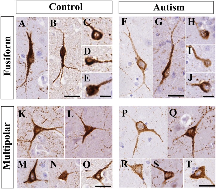
Figure 3.
The morphology of neuropeptide Y+ neurons in the caudate nucleus was similar in controls and subjects with autism. Images were taken from n = 5 controls and n = 5 patients with autism. (A–E) Fusiform neuropeptide Y+ neurons in controls. (F–J) Fusiform neuropeptide Y+ neurons in subjects with autism. (K–O) Multipolar neuropeptide Y+ neurons in controls. (P–T) Multipolar neuropeptide Y+ neurons in autism spectrum disorder. Scale bars in C–E, H–J = 10 µm, otherwise 30 µm.

