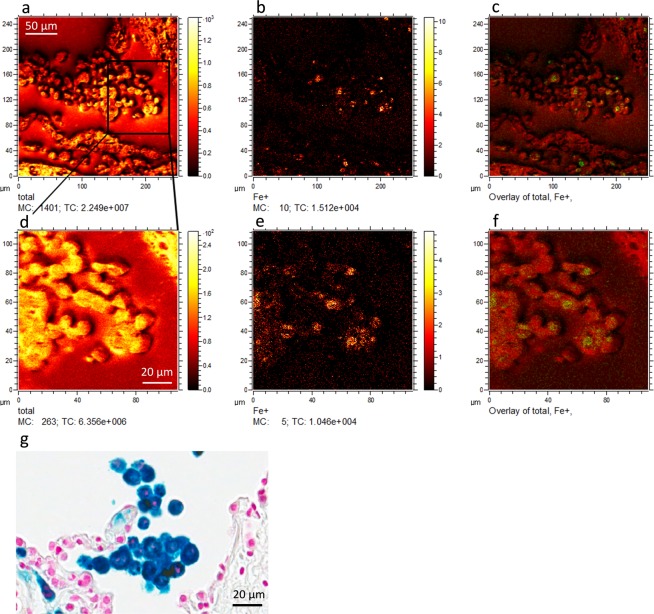
Figure 2.
Heterogeneous iron distribution has been shown in iron positive cells of COPD (GOLD IV) patients’ lung tissue. Top panel shows representative (a) total ion image, (b) the iron ion signal image, and (c) the overlay of the two images obtained from COPD (GOLD IV) patients’ lung tissue using delayed extraction mode in ToF-SIMS instrument. Bottom panel shows a region of image (a) further magnified and their corresponding (d) total ion image, (e) iron ion signal image and (f) the overlay of both. Figure 2g shows a consecutive section of Perls’ Prussian blue/ nuclear fast red stained COPD (GOLD IV) patient lung tissue including alveolar macrophages (tentatively identified). Scale bar for figure (a–c) 50µm and figure (d–g) 20µm.

