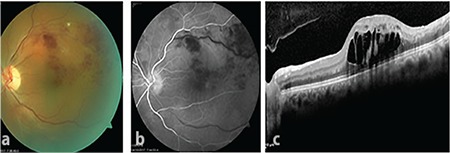
Figure 1.

At presentation, a) color fundus image showed superotemporal branch retinal vein occlusion; b) fundus fluorescein angiography showed late filling and dilation of the superotemporal vein and areas of capillary nonperfusion; and c) optical coherence tomography showed macular thickening and cystoid edema
