
Figure 3.
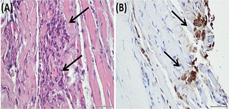
(A) Hematoxylin-eosin stain. The biopsy specimen of the right medial rectus muscle showing tumor cells (arrows) infiltrating muscle fibers; 400x. (B) Cytokeratin 7 immunohistochemistry of the specimen showing tumor cells (arrows); 400x
Official websites use .gov
A
.gov website belongs to an official
government organization in the United States.
Secure .gov websites use HTTPS
A lock (
) or https:// means you've safely
connected to the .gov website. Share sensitive
information only on official, secure websites.
(A) Hematoxylin-eosin stain. The biopsy specimen of the right medial rectus muscle showing tumor cells (arrows) infiltrating muscle fibers; 400x. (B) Cytokeratin 7 immunohistochemistry of the specimen showing tumor cells (arrows); 400x