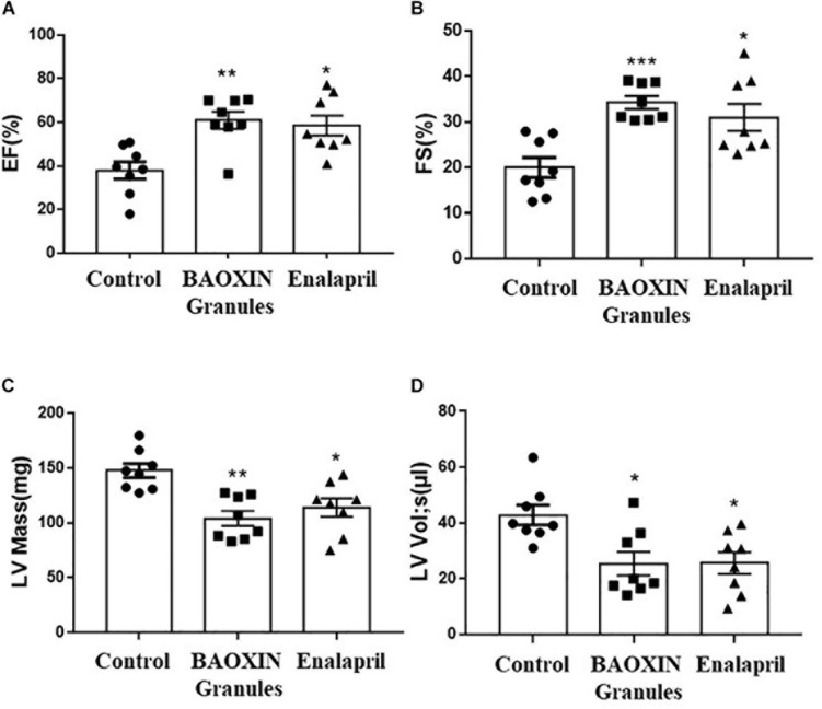
FIGURE 2.
Echocardiographic analysis of cardiac function after 4 weeks treatments. LV systolic function was reflected by quantification of EF (A), FS (B), LV Mass (C), LV Vol;s (D). The data were showed as mean and standard error of the mean (mean ± SEM). *p < 0.05 or ∗∗p < 0.01 compared with Control group, n = 8.

