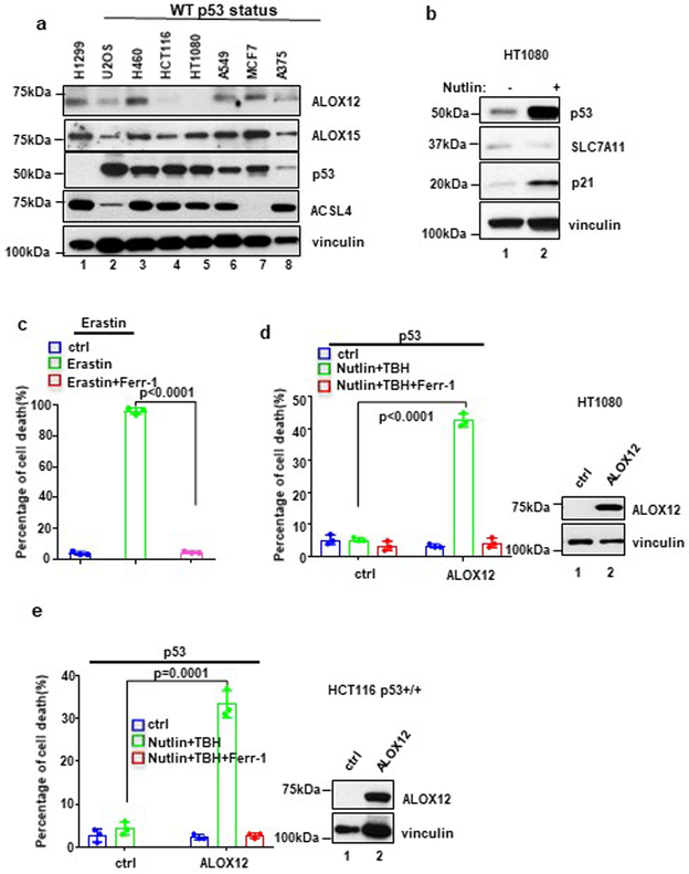
Figure 7. Mechanistic insight into p53-mediated ferroptosis.
a) Western blot analysis of a panel of cancer cell lines. The experiments were repeated twice, independently, with similar results.
b) Western blot analysis of HT1080 cells with or without Nutlin (10uM) treatment. The experiments were repeated twice, independently, with similar results.
c) HT1080 cells treated with Erastin (10uM) and Ferr-1 (2uM) treatment for 8h. Quantification of cell death is shown. Error bars are mean ± s.d., n=3 independent experiments.
d) HT1080 cells transfected with ALOX12 were pre-incubated with Nutlin (10uM) for 12h, then treated with TBH (200uM), Nutlin (10uM) and Ferr-1 (2uM) for 8h. Quantification of cell death is shown. Western blot analysis of HT1080 cells transfected with ALOX12. Error bars are mean ± s.d., n=3 independent experiments. The Western blot experiments were repeated twice, independently, with similar results.
e) HCT116 p53+/+ cells were pre-incubated with Nutlin (10uM) for 12h, then treated with Nutlin (10uM), TBH (200uM), and Ferr-1 (2uM) as indicated for 12h. Quantification of cell death is shown. Western blot analysis of HCT116 p53+/+ cells transfected with ALOX12. Error bars are mean ± s.d., n=3 independent experiments. All P values (c,d,e) were calculated using two-tailed unpaired Student’s t-test. Detailed statistical tests are described in the Methods. The Western blot experiments were repeated twice, independently, with similar results. Scanned images of unprocessed blots are shown in Supplementary Fig. 9. Raw data are provided in Supplementary Table 1.

