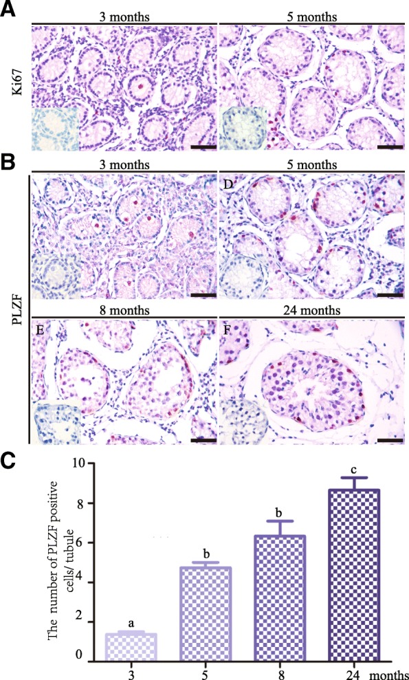
Fig. 2.

Immunohistological analyses of germ cell differentiation in the yak testis at four different developmental stages. a Immunostaining for Ki67 in testicular sections from yaks at 3 and 5 months. b Immunostaining for PLZF in testicular sections from yaks at 3, 5, 8 and 24 months. c Quantification of PLZF+ cells per seminiferous tubules at four different developmental stages. Negative control was shown in the lower-left corner. Different letter denotes significantly different at P < 0.05. Scale bar = 50 μm
