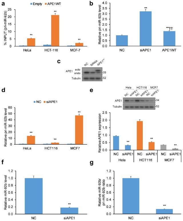
Figure 1.
APE1 binds to pri-miR-92b. (a) Validation of APE1-pri-miR-92b binding in the HeLa, HCT-116, and MCF-7 cancer cell lines. qRT-PCR of APE1-bound pri-miR-92b in cell lines transfected with APE1 WT FLAG-tag protein vector or empty vector. Data reported as proportion of immunoprecipitated pri-miR-92b relative to pri-miR-92b in total inputted RNA. (b) Pri-miR-92b levels evaluated in APE1-silenced HeLa cells. (c) Western blotting validation of APE1 silencing in HeLa cells with tubulin normalization. (d) Pri-miR-92b levels in APE1-silenced HeLa, HCT-116, and MCF-7 cell lines. (e) Western blotting validation of APE1 silencing in HeLa, HCT-116, and MCF-7 cells with tubulin normalization. (f) Mature miR-92b levels and (g) mature miR-92b-to-pri-miR ratios in APE1-silenced HeLa cells. Mature miR-92b levels were normalized to RNU44, while pri-miR-92b levels were normalized to GAPDH.
Data are represented as means ± SEMs. *p < 0.05, **p < 0.01.
APE1, apurinic/apyrimidinic endodeoxyribonuclease 1; GAPDH, glyceraldehyde 3-phosphate dehydrogenase; qRT-PCR, quantitative reverse transcription polymerase chain reaction; SEM, standard error of the mean; WT, wild type.

