. 2019 Jun 5;10(27):6651–6660. doi: 10.1039/c9sc01441a

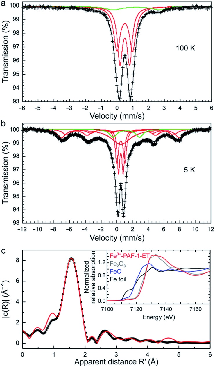
Fig. 4. Iron-57 Mössbauer spectra of PAF-1–ET obtained at (a) 100 and (b) 5 K. Red and green fits were assigned to iron(iii) (91%) and iron(ii) (9%), respectively. The total fit is shown in black. (c) The Fourier transform of the k3-weighted iron K-edge EXAFS data of iron(iii) adsorbed in PAF-1–ET (black dots); the best two-shell fit (red curve) was achieved with an immediate coordination environment of six O atoms at a distance of 2.00(1) Å and 12 C atoms at a distance of 3.06(4) Å; (inset) room-temperature XANES spectra obtained at the iron K-edge for PAF-1–ET (red curve) and Fe2O3, FeO, and Fe foil (gray, blue, and black curves, respectively).

Fig. 4