
Fig. 3.
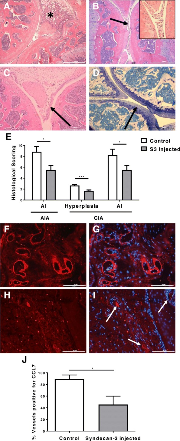
Therapeutic effects of syndecan-3 in the antigen-induced arthritis (AIA) and collagen-induced arthritis (CIA) mouse models. Histological sections were analysed for any effects on inflammation and tissue damage after a syndecan-3 intra-articular injection. Parameters measured for AIA were a thickening of the synovium*, b exudate in the joint space (arrow) with insert showing higher magnification, c hyperplasia of the synovial lining layer (arrow) and d loss of proteoglycans from articular cartilage (arrow showing loss of toluidine blue staining). The histological scores for 3 days post-injection are displayed in e (means and standard errors are shown, n = 4 mice in each group for AIA and n = 10 mice for CIA; *P < 0.05; *** P < 0.001; AI, arthritis index). f, g Blood vessels positive for CCL7 in the control (PBS injected) AIA group. h, i Blood vessels negative for CCL7 staining in the test (syndecan-3 injected) AIA group, as indicated by arrows. g, i Merged DAPI images. j Five random fields of view per mouse scored based on the percentage of the blood vessels positive for CCL7 staining on synovial endothelial cells after PBS (control) or syndecan-3 intra-articular injection (data are means with standard errors, n = 7 AIA mice per group; *P < 0.05). The magnification bars in a–d represent 200 μm and f–i 50μm