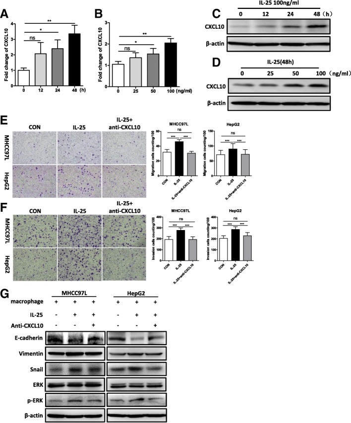
Fig. 3.
IL-25-induced M2 macrophages promote the EMT process in HCC cells via CXCL10. a-d Macrophages (derived from THP-1) were treated with IL-25 in a time- and concentration-dependent manner. a and b CXCL10 gene expression was quantified by RT-qPCR. c and d CXCL10 protein level was determined by Western blotting. e-g HCC cells were co-cultured with M0 or M2 macrophages. Then, anti-CXCL10 antibody was added to the M2 macrophage culture medium to neutralize CXCL10 protein. Migration (e) and invasion (f) of HCC cells were determined by Transwell assay. Statistical data were shown at the right. Bar, 100 μm. g Mesenchymal maker vimentin, EMT regulator Snail, epithelial marker E-cadherin, extracellular signal-regulated kinase (ERK), and p-ERK were detected by Western blotting. *p < 0.05, **p < 0.01, ***p < 0.001, ns, no significance

