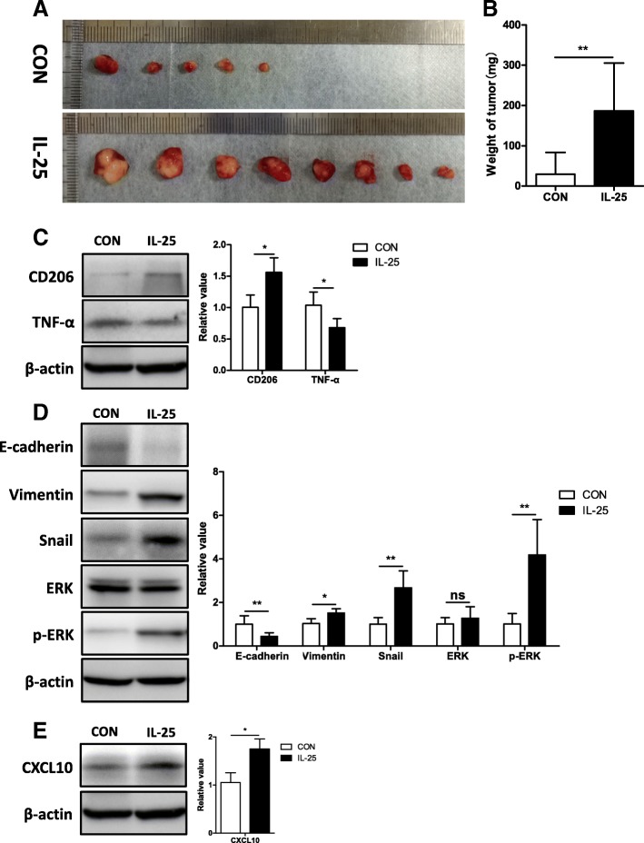
Fig. 4.
IL-25-induced M2 macrophages promote tumorigenesis and EMT of HCC cells in vivo. a Images of tumors from each group. b Tumor weight at the time of sacrifice. c Tumor necrosis factor-α (TNF-α) and CD206 in the tumor tissue of each group were detected by Western blotting. Statistical data were shown at the right. d Mesenchymal maker vimentin, EMT regulator Snail, epithelial marker E-cadherin, extracellular signal-regulated kinase (ERK), and p-ERK were detected in the tumor tissue of each group by Western blotting. Statistical data were shown at the right. e Chemokine CXCL10 was detected in the tumor tissue of each group by Western blotting. Statistical data were shown at the right. *p < 0.05, **p < 0.01, ns, no significance

