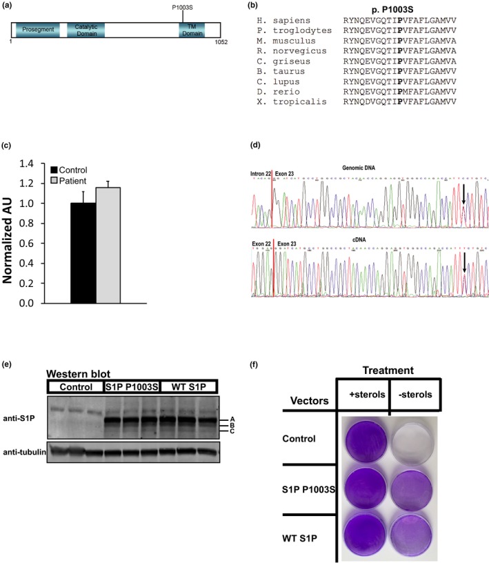
Figure 1.

Genetic and functional analysis of S1P Pro1003Ser. (a) Schematic of S1P protein with the Pro1003Ser mutation and S1P protein domains indicated, TM, transmembrane domain. (b) Multispecies alignment of S1P amino acid sequences demonstrating the mutated proline 1003 residue is conserved (shown in bold). (c) mRNA expression levels of the MBTPS1 transcript in cultured control‐ and patient‐derived skin fibroblasts. n = 3. (d) Sanger sequencing trace files of patient genomic DNA (top) and cDNA (bottom)‐derived PCR products at the MBTPS1 c.3007C>T locus. Intron–exon boundary (genomic) and exon–exon junction (cDNA) are indicated. Arrows denote the variant location. (e) Western blot of whole‐cell lysates (60μg) from SRD‐12B cells transiently transfected with mock, WT S1P, or S1P Pro1003Ser plasmids after 24 hr. S1P‐A, B, and C forms are indicated. Blots were probed with S1P and α‐tubulin (loading control) antibodies. n = 3. (f) SRD‐12B cells were transfected as in (e) and grown either in medium supplemented with lipids and cholesterol (left column) or in lipid‐ and cholesterol‐free medium (right column) for 7 days followed by fixation in methanol and crystal violet staining, as reported previously (Rawson et al., 1998). Images are representative of three independent experiments
