Figure 1.

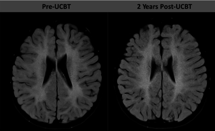
Representative axial FLAIR sequences from brain MRI. Left MRI was performed at age 4 years, 8 months (pre‐UCBT) and at age 6 years, 8 months (2 years post‐UCBT) in a boy with β‐mannosidosis

Representative axial FLAIR sequences from brain MRI. Left MRI was performed at age 4 years, 8 months (pre‐UCBT) and at age 6 years, 8 months (2 years post‐UCBT) in a boy with β‐mannosidosis