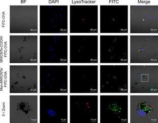
Figure 9.

Confocal images of BMDCs incubated with FITC‐labeled OVA, MWCNTs‐COOH/FITC‐OVA, and Man‐MWCNTs/FITC‐OVA for 4 h. Later endosomes and lysosomes (red) were stained with lysoTacker‐Red, while OVA (green) were prelabeled with FITC. Nuclei (blue) were stained with DAPI.
