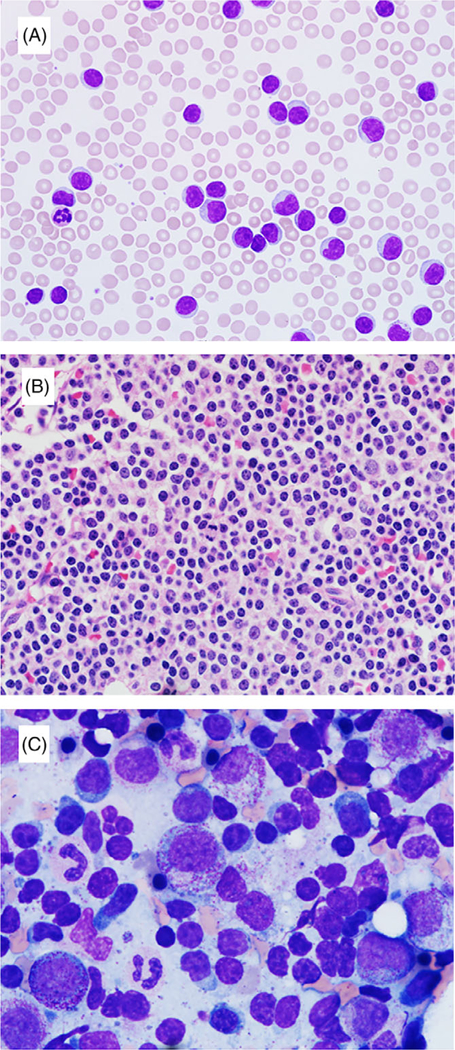
FIGURE 1.

Morphologic findings in CLL patients with IGH-BCL3 translocation: A peripheral blood (A; 600×) and a core biopsy (B; 600×) from a same case show mostly small-sized lymphocytes with nuclear irregularity, modest cytoplasm, and some with single, small nucleolus. A bone marrow aspirate smear (C; 1000×) from another case shows a subset of cells with slightly irregular nuclear contours, moderate basophilic cytoplasm, and plasmacytoid appearance
