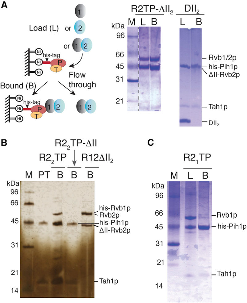
Figure 4. Biochemical Verification of the Importance of Insertion Domain in ScR2TP Assembly.

Solid ovals represent Rvb1p (1), Rvb2p, or Rvb2p DII deletion (2), Pih1p (P), and Tah1p (T). SDS-PAGE gels are stained by either Coomassie brilliant blue (blue) or silver nitrate (yellow). Samples analyzed on gels are denoted as L for load, B for bound, PT for Pih1p-Tah1p control, and M for protein markers.
(A) Schematic representation of the assembly and pull-down method (left) and SDS-PAGE gel analysis of Pih1p-Tah1p binding to Rvb1/2p lacking domain II in Rvb2p (ScR2TP-ΔII2) or DII2 (domain II of Rvb2p) (right).
(B) SDS-PAGE gel (silver-stained), showing Pih1p-Tah1p binding to Rvb2p (R22TP) but not to Rvb2p lacking its domain II (R22TP-ΔII). The last lane (R12-ΔII2) shows binding of Rvb1p to the same Rvb2p lacking its domain II (Rvb2p-ΔII) sample used for the R22TP-ΔII complex formation.
(C) SDS-PAGE gel, showing formation of the Pih1p-Tah1p-Rvb1p complex (R1TP).
