Figure 2.
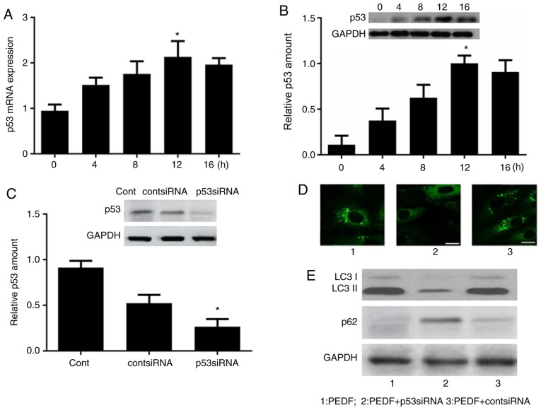
p53 is critical for PEDF-induced autophagy. Human umbilical vein endothelial cells were treated with PEDF for 0, 4, 8, 12 and 16 h. (A) p53 mRNA and (B) protein expression was detected by reverse transcription-quantitative polymerase chain reaction and western blot analysis. *P<0.05 vs. 0 h. (C) p53 siRNA was established and western blot analysis was performed to confirm the effect of p53 gene silencing. *P<0.05 vs. control group. (D) Fluorescence microscopy of green fluorescent protein-positive autophagosomes in the PEDF, PEDF + p53 siRNA and PEDF + cont siRNA groups. Scale bar, 10 µm. (E) Western blot analysis in the PEDF, PEDF + p53 siRNA and PEDF + cont siRNA groups. LC3, microtubule-associated protein light chain 3; PEDF, pigment epithelium-derived factor; si, small interfering.

