Figure 2.
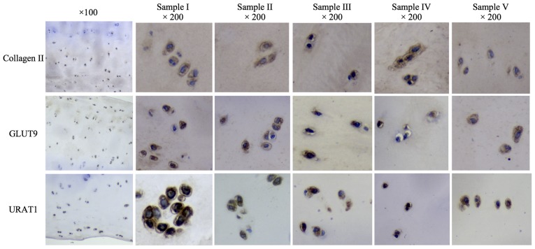
Histological analysis of urate transporter expression in human cartilage. Human articular cartilage samples (I to V) exhibited positive expression of collagen II, GLUT9 and URAT1, as determined via immunohistochemical analysis. Digital images were captured using a Nikon Microphot-FX microscope [magnification, ×100 (Sample II) and ×200 (all five samples)]. Representative images are shown. GLUT9, glucose transporter 9; URAT1, urate transporter 1.

