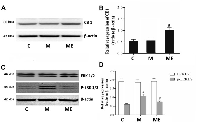
Figure 3.
Effects of EA on CB1, ERK1/2 and p-ERK1/2 expression in morphine-induced hyperalgesia. Spinal cord tissue from different groups was collected 8 days after intrathecal treatment with saline, morphine and EA. (A) CB1, (C) ERK1/2 and p-ERK1/2 levels were detected by western blotting. Quantitative analysis of (B) CB1, (D) ERK1/2 and p-ERK1/2 are shown as the ratio of protein relative density to β-actin. Data are expressed as the mean ± standard deviation (n=6 rats/group). *P<0.05 vs. the C group, #P<0.05 vs. the M group. EA, electroacupuncture; CB1, cannabinoid receptor 1; ERK1/2, extracellular signal-regulated kinase 1/2; p, phosphorylated; C, control; M, chronic morphine; ME, morphine + EA at ST36-GB34.

