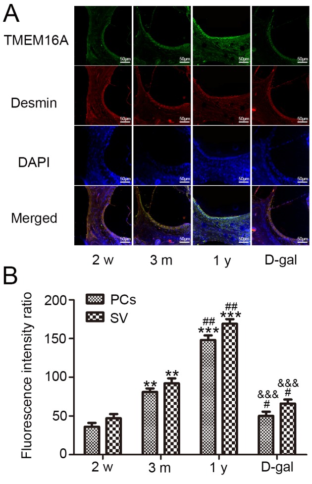
Figure 6.

Distribution of TMEM16A in the PCs in the SV of guinea pigs. (A) SV was stained for TMEM16A (green) and desmin (red) as a specific marker of PCs (arrows indicate PCs in the SV). DAPI (blue) was used to stain nuclei. Scale bars=50 µm. (B) Quantification of TMEM16A expression at different ages; similar fluorescence intensity was observed in PCs and the overall SV. Data are presented as the mean ± standard deviation (n=10). **P<0.01, ***P<0.001 vs. 2 weeks; #P<0.05, ##P<0.01 vs. 3 months; &&&P<0.001 vs. 1 year. D-gal, D-galactose; TMEM16A, transmembrane protein 16; SV, stria vascularis; PC, pericytes.
