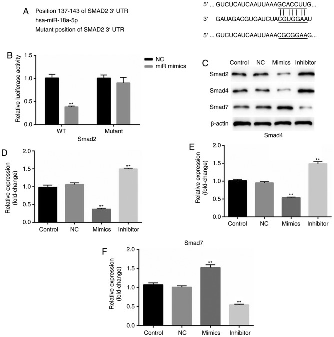
Figure 5.
miR-18a-5p directly targets Smad2. (A) Binding sites between the 3′-UTR of Smad2 and miR-18a-5p. (B) A dual-luciferase reporter assay was used to detect the luciferase activity. **P<0.01 vs. NC. (C) Protein expression levels of Smad2, Smad4 and Smad7 were examined by western blotting in SCC9 cells. (D-F) mRNA levels of Smad2, Smad4 and Smad7 were assessed by reverse transcription-quantitative polymerase chain reaction. **P<0.01 vs. the control. 3′-UTR, 3′-untranslated region; miR-18a-5p, microRNA-15a-5p; NC, negative control.

